Digitization of the Last European Elephant
3D Digitization and Printing of the Last European Elephant
This project brings engineering tools into palaeontology by digitizing, modelling and 3D printing skeletal remains of Palaeoloxodon tiliensis, the dwarf elephant from Tilos considered the last European elephant. By combining CT scanning, laser scanning, CAD modelling, allometric analysis and additive manufacturing, the work creates accurate digital and physical replicas for research, comparison, exhibition and education.
Project Scope and Palaeontological Challenge
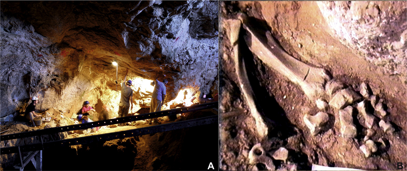
Charkadio Cave on Tilos Island preserves an exceptional fossil record of the insular dwarf elephant Palaeoloxodon tiliensis. The material is scientifically valuable but incomplete: many skeletal elements are isolated, fragmented or represented by different ontogenetic stages, and no complete adult articulated skeleton is available.
The project addresses this challenge by combining palaeontological data with engineering workflows. Fossil dimensions, taphonomical observations, allometric relationships and 3D scan data are integrated so missing measurements can be estimated and skeletal elements can be reconstructed at appropriate relative proportions.
MD-Lab’s Contribution
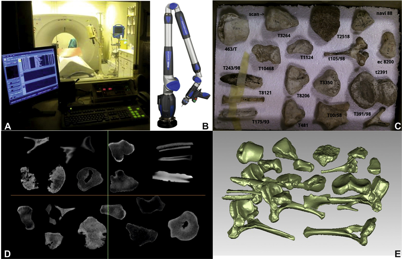
MD-Lab’s engineering contribution centers on digital reconstruction and rapid prototyping. The work translates fragile fossil material into high-quality digital geometry using CT data, laser scanning, point-cloud processing, CAD modelling and surface reconstruction.
The resulting CAD models are resized, mirrored where needed, prepared as STEP files and fabricated with additive manufacturing technologies. This makes it possible to study, compare, exhibit and exchange fossil replicas while reducing handling risks for the original specimens.
Fossil Context and Material
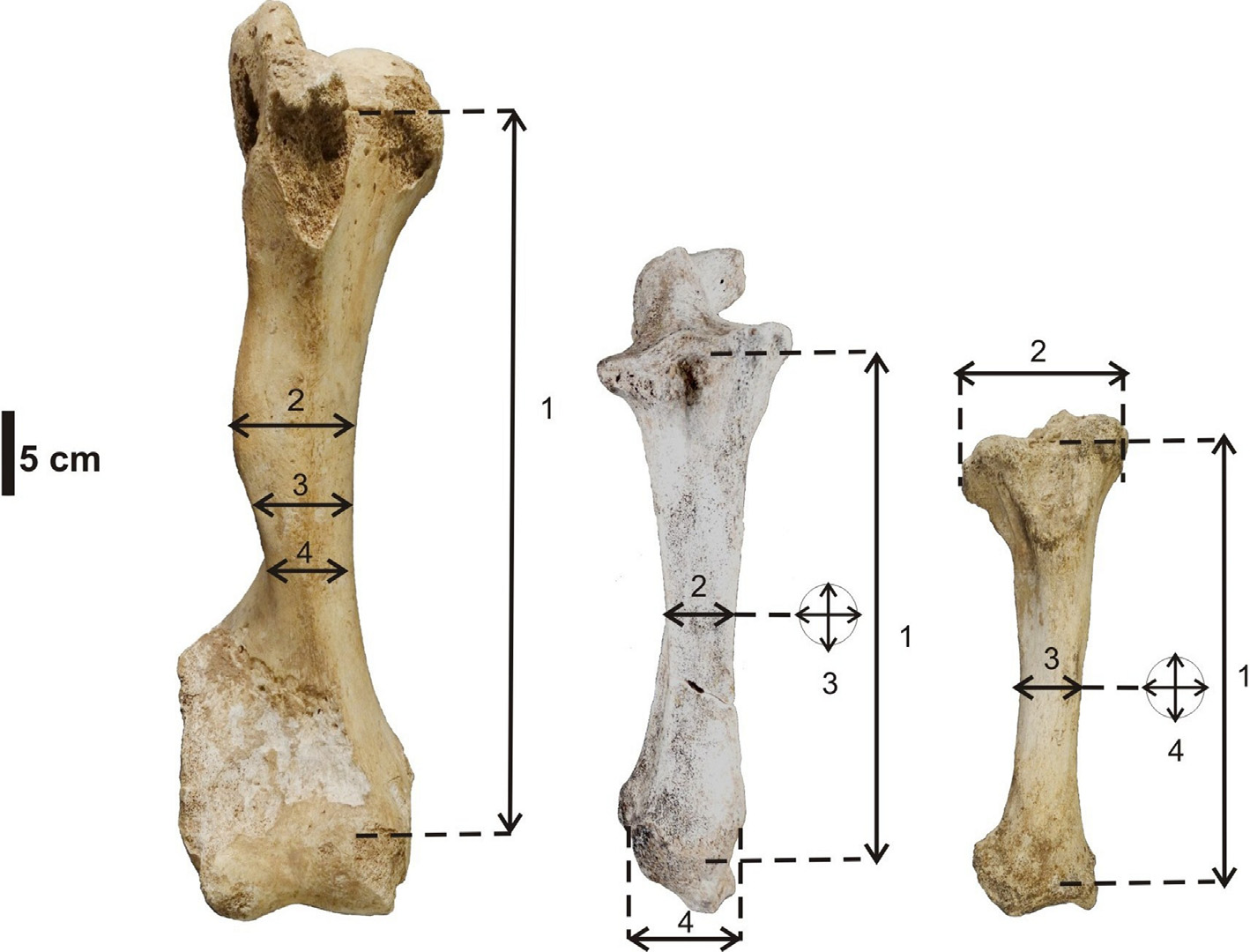
The studied material comes from Charkadio Cave, one of the richest Mediterranean sites for dwarf elephant remains. The available specimens include humerus, ulna, tibia and other skeletal elements from infant, juvenile and adult individuals.
Because the fossil record is incomplete, the project does not simply copy existing bones. It uses anatomical association, measurements and allometric scaling to establish realistic proportions before digital models are finalized and printed.
Allometry and Dimensional Reconstruction
Statistical and allometric analyses were used to estimate the dimensions of adult skeletal elements. The methodology relates available measurements across comparable bones and ontogenetic stages so that representative adult proportions can be reconstructed.
This step is essential because scanning alone cannot solve missing geometry. The final digital models depend on both captured surface morphology and mathematically supported dimensional adjustment.
CT Scanning, Laser Scanning and CAD Modelling
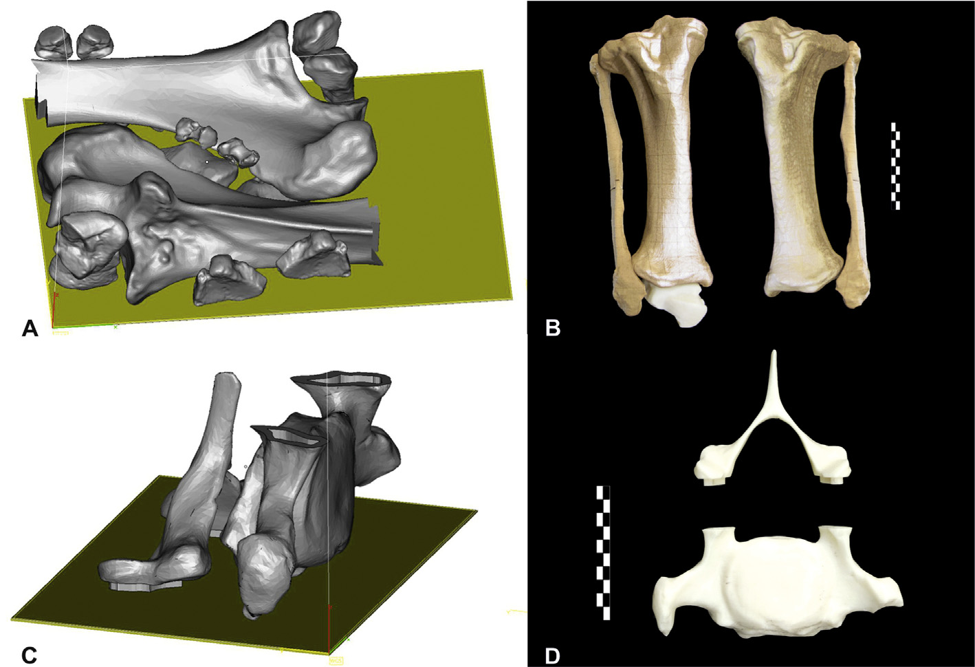
Two complementary digitization routes were used. Computed tomography captured groups of smaller specimens and internal geometry, while laser scanning provided surface data for fragile or larger elements that could be processed as point clouds.
The raw scan data was converted into watertight digital surfaces and then into CAD models. These models could be smoothed, mirrored, dimensionally adjusted and prepared for additive manufacturing without exposing the original fossils to repeated handling.
Digital Morphological Comparison
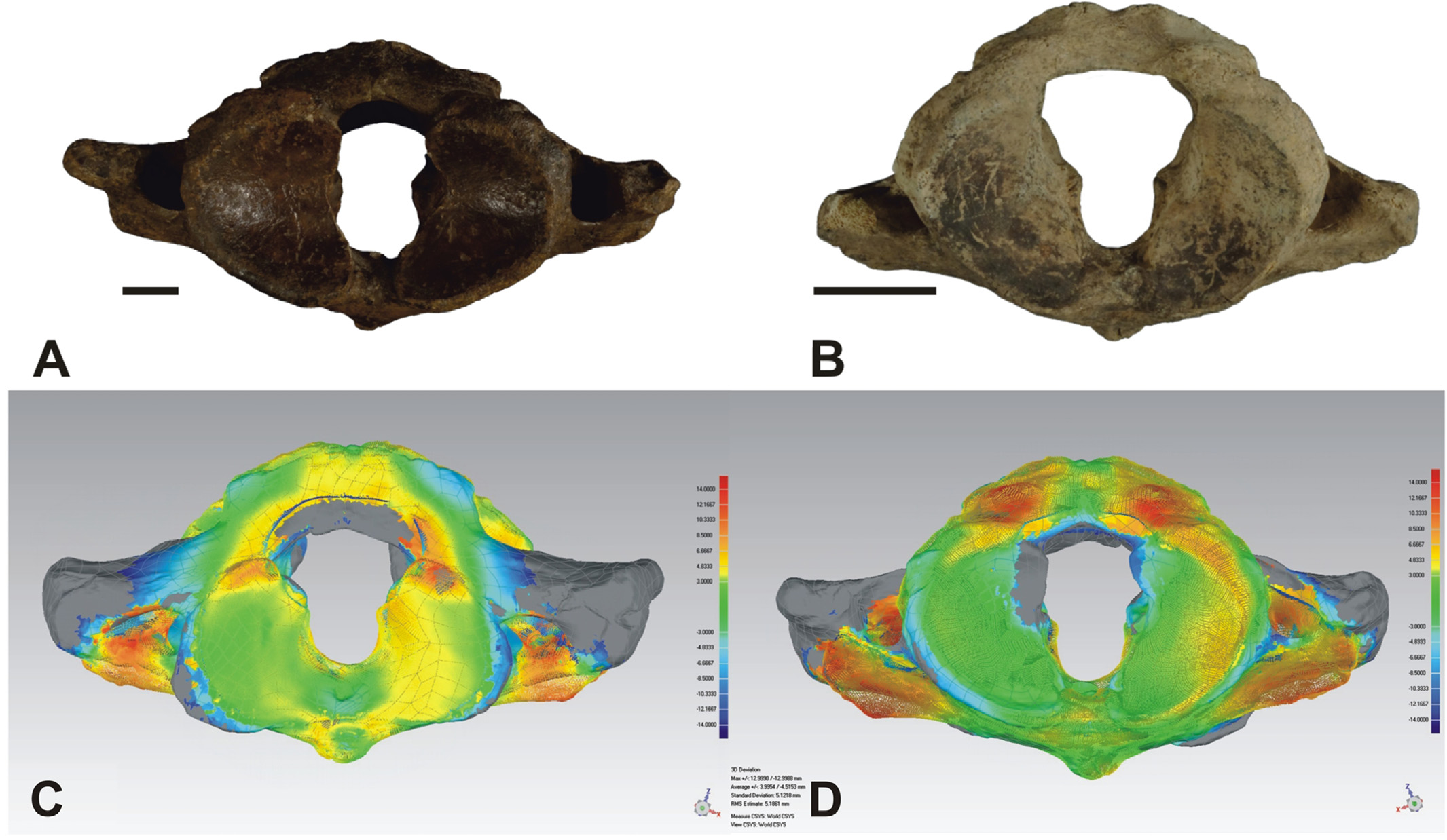
The digital models also support comparative palaeontology. In the atlas comparison, 3D models of Palaeoloxodon antiquus and Palaeoloxodon tiliensis were aligned and compared through a color-scale deviation map.
This approach makes morphological differences easier to quantify and communicate. Instead of relying only on visual inspection, the model highlights regions of similarity and difference across the matched fossil surfaces.
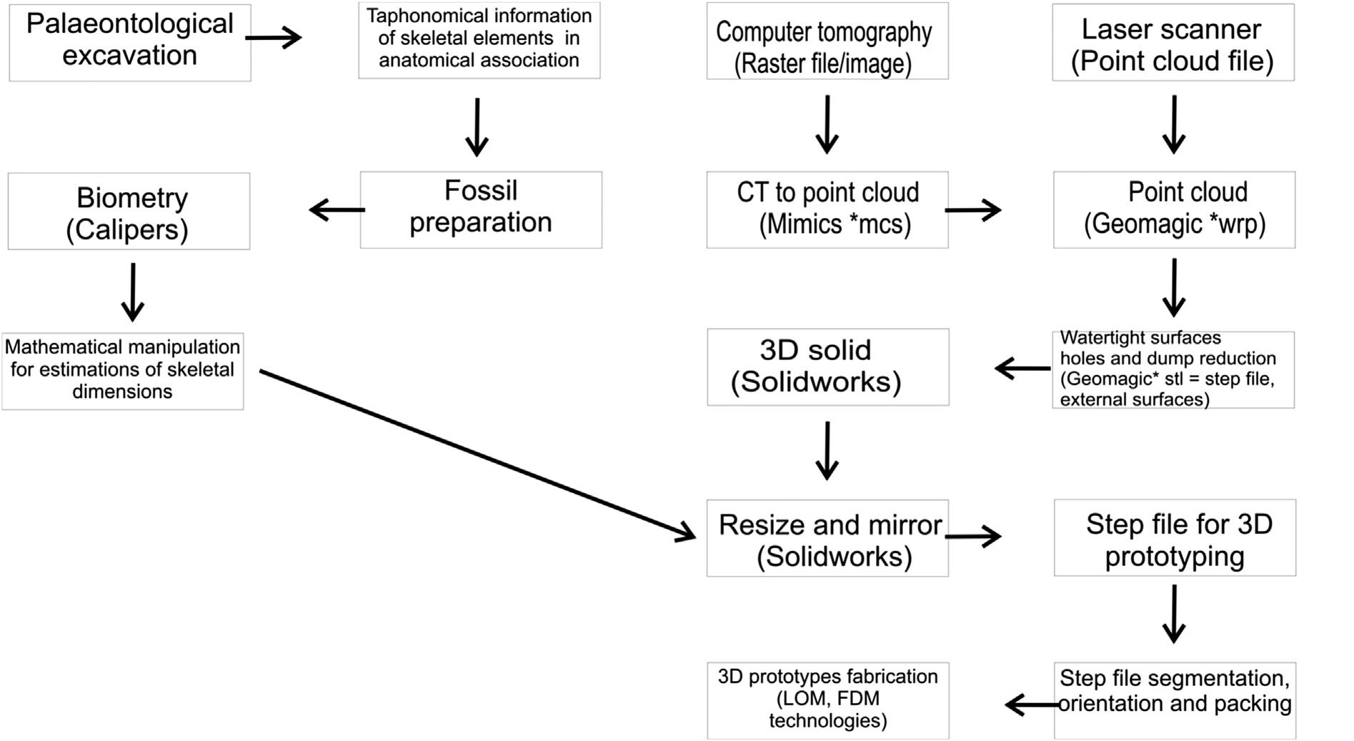
From Fossil Excavation to 3D Printed Replica
The project establishes a complete workflow: excavation and taphonomical recording, fossil preparation, biometrical measurement, CT or laser scanning, point-cloud processing, CAD modelling, dimensional correction, STEP export, build preparation and fabrication.
This workflow is important because it is transferable. It can be applied to other palaeontological collections where original specimens are incomplete, fragile or too valuable to move frequently between institutions.
Main Outcomes
More than forty skeletal elements were digitally modelled and printed, using LOM and FDM additive manufacturing according to the size, weight and fabrication needs of each replica. The process enabled mirrored bones, reconstructed missing parts and dimensionally consistent skeletal elements.
The project also created a research-ready digital model base for Palaeoloxodon tiliensis. These data can support scientific comparison, educational use, museum display and inter-institutional exchange without risking damage to irreplaceable fossils.
Engineering Significance
The work is significant because it shows how mechanical engineering methods can extend palaeontological research. CT scanning, laser scanning, CAD modelling and rapid prototyping do not replace the fossil record; they make it more accessible, measurable and shareable.
By creating accurate digital and physical replicas of the Tilos dwarf elephant, the project supports virtual collections, safer study of fragile material, morphological comparison between taxa and more engaging educational exhibits for museums, universities and schools.